

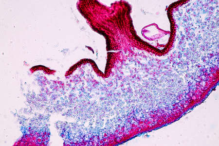
Bowen's Disease Tumor under the microscope 100x
Коллекция по умолчанию
Коллекция по умолчанию
Создать новую
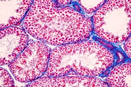
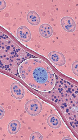
Anatomy and Histological Epididymis and Testis human cells under microscope.
Коллекция по умолчанию
Коллекция по умолчанию
Создать новую
Histopathology of alveoli, light micrograph, photo under microscope
Коллекция по умолчанию
Коллекция по умолчанию
Создать новую
Pancreas cancer, light micrograph, photo under microscope
Коллекция по умолчанию
Коллекция по умолчанию
Создать новую

Feeler snail's eye tissue under the microscope 200x
Коллекция по умолчанию
Коллекция по умолчанию
Создать новую

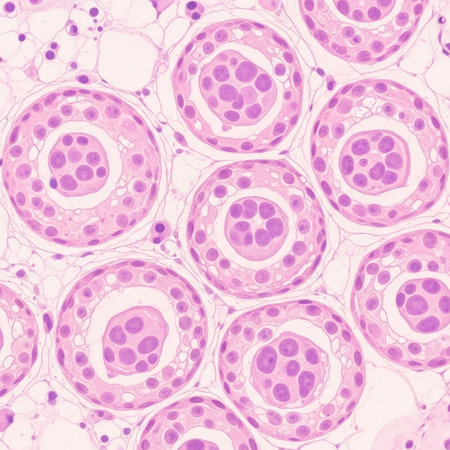
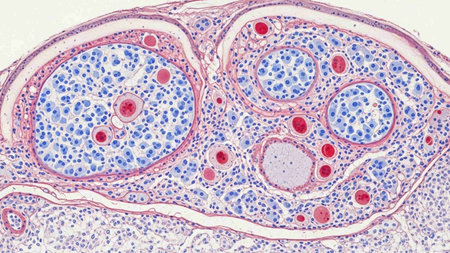
Cross section Human testis under microscope view for education histology, Human tissue
Коллекция по умолчанию
Коллекция по умолчанию
Создать новую

Reproductive parts: stigma, style, stamens, filament, petal. Structure floral- male reproductive organ of the flower of angiosperms
Коллекция по умолчанию
Коллекция по умолчанию
Создать новую
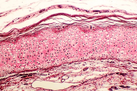
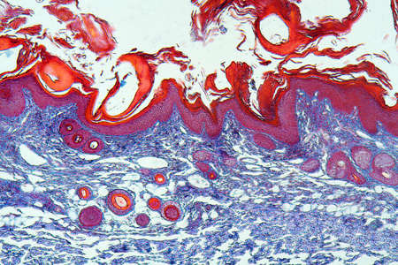
Cross section of a human skin under microscope view for education in laboratory.
Коллекция по умолчанию
Коллекция по умолчанию
Создать новую
Photomicrograph showing histological features of benign prostatic hyperplasia. Enlarged prostate gland with nodular proliferation of glandular and stromal components.
Коллекция по умолчанию
Коллекция по умолчанию
Создать новую
Heather leaf cross section under the microscope, 200x
Коллекция по умолчанию
Коллекция по умолчанию
Создать новую
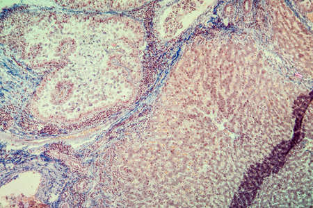
Histopathology of cirrhosis under the microscope 100x.
Коллекция по умолчанию
Коллекция по умолчанию
Создать новую
Cell
Коллекция по умолчанию
Коллекция по умолчанию
Создать новую
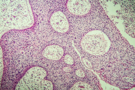
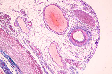
Light micrograph of ovary showing primordial, primary and secondary follicules. Light microscopy, hematoxylin and eosin stain, magnification 200x
Коллекция по умолчанию
Коллекция по умолчанию
Создать новую
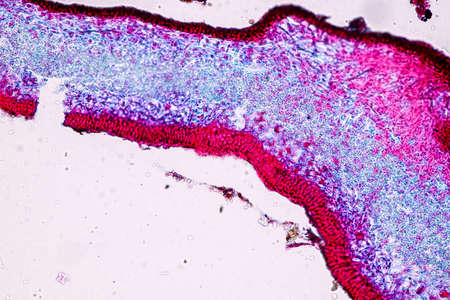
Bowen's Disease Tumor under the microscope 100x
Коллекция по умолчанию
Коллекция по умолчанию
Создать новую
Anatomy and Histological Ovary, Testis and Sperm human cells under microscope.
Коллекция по умолчанию
Коллекция по умолчанию
Создать новую
Microscopic Anatomy of Lycoperdon Bovista Puffball Gleys and Spore Structure
Коллекция по умолчанию
Коллекция по умолчанию
Создать новую
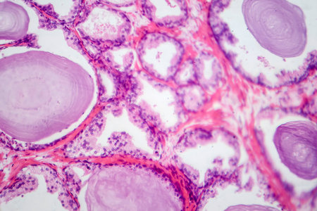
Histopathology of prostate gland hyperplasia, light micrograph, photo under microscope
Коллекция по умолчанию
Коллекция по умолчанию
Создать новую
Anatomy and Histological Epididymis and Testis human cells under microscope.
Коллекция по умолчанию
Коллекция по умолчанию
Создать новую
Condyloma acuminatum, also known as genital warts. Light micrograph, photo under microscope
Коллекция по умолчанию
Коллекция по умолчанию
Создать новую

Cell- science background. Esophagus of the dog- cross section
Коллекция по умолчанию
Коллекция по умолчанию
Создать новую
Club moss flower with seeds under the microscope 100x
Коллекция по умолчанию
Коллекция по умолчанию
Создать новую
Coccidiosis of liver tissue under the microscope 100x
Коллекция по умолчанию
Коллекция по умолчанию
Создать новую
Histopathology of human kidney cross section under microscope for education.
Коллекция по умолчанию
Коллекция по умолчанию
Создать новую
Photomicrograph showing histological features of benign prostatic hyperplasia. Enlarged prostate gland with nodular proliferation of glandular and stromal components.
Коллекция по умолчанию
Коллекция по умолчанию
Создать новую

Anatomy and Histological Bone, Elastic cartilage human and Joint of human foetus under the microscope for education.
Коллекция по умолчанию
Коллекция по умолчанию
Создать новую

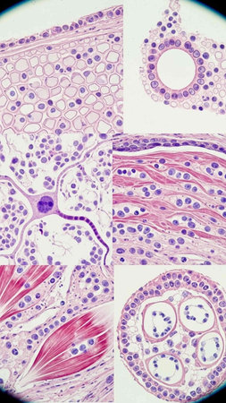
The study of tissue samples of Trachea of Cat, Epididymis, Prostate, Uterus with embryo of rat and Mammary gland cow under the microscope in Lab.
Коллекция по умолчанию
Коллекция по умолчанию
Создать новую
Cell Gene Microscopic Series
Коллекция по умолчанию
Коллекция по умолчанию
Создать новую
Scalp and hair follicles of human under the microscope in Lab.
Коллекция по умолчанию
Коллекция по умолчанию
Создать новую

Histological Uterus human, Uterine tube human, Placenta human and Umbilical cord Human under the microscope for education
Коллекция по умолчанию
Коллекция по умолчанию
Создать новую
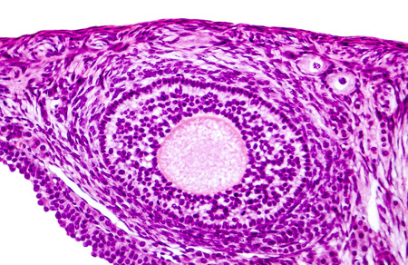
Ovarian follicles. Light microscopy, hematoxylin and eosin stain, magnification 200x. Colors are enhanced for better visualisation
Коллекция по умолчанию
Коллекция по умолчанию
Создать новую
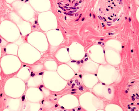
Small adipocyte lobule located in a connective tissue. A small nerve is located in the upper right corner. Light micrograph. H&E stain.
Коллекция по умолчанию
Коллекция по умолчанию
Создать новую
Anatomy and Histological Epididymis and Testis human cells under microscope.
Коллекция по умолчанию
Коллекция по умолчанию
Создать новую
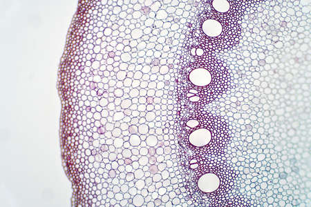
Root plant show root vascular tissue under light microscope view for education.
Коллекция по умолчанию
Коллекция по умолчанию
Создать новую
Histopathology of human hair follicle, 3D illustration.
Коллекция по умолчанию
Коллекция по умолчанию
Создать новую
Showing Light micrograph of the Thyroid gland and Thymus gland human Child under the microscope for education in the laboratory.
Коллекция по умолчанию
Коллекция по умолчанию
Создать новую
Showing Light micrograph of the Adrenal gland and Urinary bladder human under the microscope for education in the laboratory.
Коллекция по умолчанию
Коллекция по умолчанию
Создать новую

Anatomy and Histological Epididymis and Testis human cells under microscope.
Коллекция по умолчанию
Коллекция по умолчанию
Создать новую
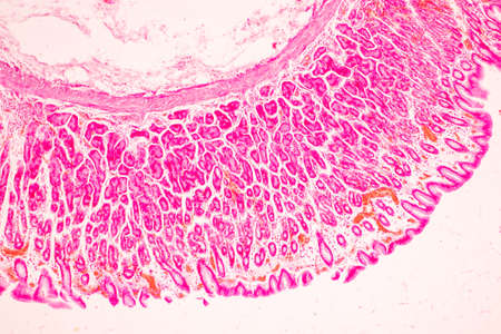
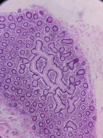
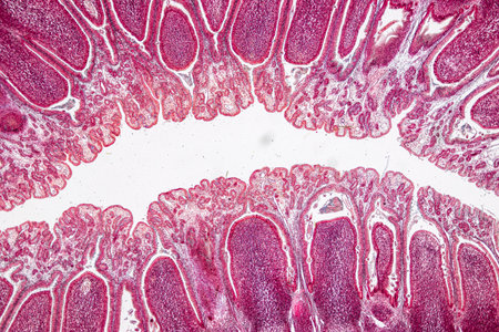
Tissue of Stomach Human under the microscope in Lab.
Коллекция по умолчанию
Коллекция по умолчанию
Создать новую
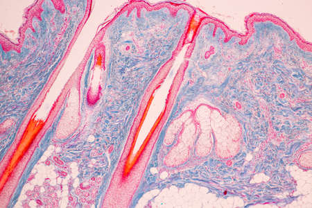
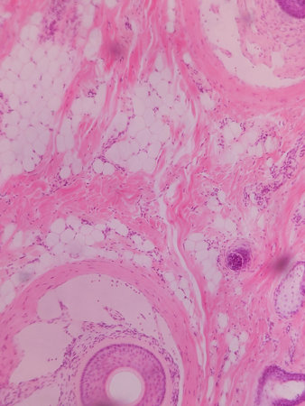
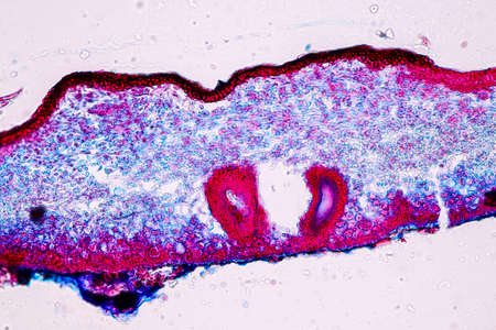
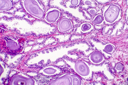
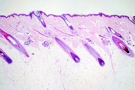
Scalp with hair roots tissue under the microscope 100x
Коллекция по умолчанию
Коллекция по умолчанию
Создать новую
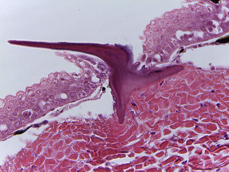
Tongue Tissue with taste buds across 200x
Коллекция по умолчанию
Коллекция по умолчанию
Создать новую
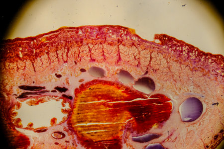
Human pancreas in the form of a cross section.
Коллекция по умолчанию
Коллекция по умолчанию
Создать новую
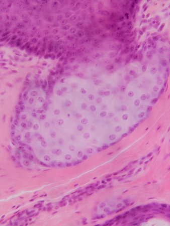
Microscopic view of tissue with pink and purple staining, showing cellular structures and connective tissue
Коллекция по умолчанию
Коллекция по умолчанию
Создать новую

Host cells with spores (mold) are inside wood under the microscope for education.
Коллекция по умолчанию
Коллекция по умолчанию
Создать новую
Education anatomy and Histological sample of Human under the microscope.
Коллекция по умолчанию
Коллекция по умолчанию
Создать новую

Host cells with spores (mold) are inside wood under the microscope for education.
Коллекция по умолчанию
Коллекция по умолчанию
Создать новую
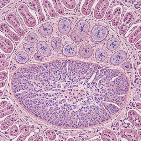
Tissue of Small intestine (Duodenum), Large intestine Human and Stomach Human under the microscope in Lab.
Коллекция по умолчанию
Коллекция по умолчанию
Создать новую
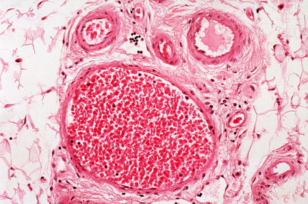
Blood vessels
Коллекция по умолчанию
Коллекция по умолчанию
Создать новую
A microscopic view of tissue with pink and purple staining, showing cellular structures and patterns
Коллекция по умолчанию
Коллекция по умолчанию
Создать новую
Photomicrograph showing histological features of benign prostatic hyperplasia. Enlarged prostate gland with nodular proliferation of glandular and stromal components.
Коллекция по умолчанию
Коллекция по умолчанию
Создать новую

Histological Uterus human, Uterine tube human, Placenta human and Umbilical cord Human under the microscope for education.
Коллекция по умолчанию
Коллекция по умолчанию
Создать новую
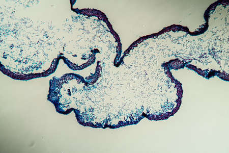
Cliated epithelium of human under the microscope in Lab.
Коллекция по умолчанию
Коллекция по умолчанию
Создать новую
Johannes berry fruit cross 100x
Коллекция по умолчанию
Коллекция по умолчанию
Создать новую

Abstract science background- pyloric division of the stomach of the dog. Cell biology
Коллекция по умолчанию
Коллекция по умолчанию
Создать новую
Histopathology of human pancreas under microscope 100x.
Коллекция по умолчанию
Коллекция по умолчанию
Создать новую
Microscopic Texture of Dogfish Shark Skin and Placoid Scale Morphology
Коллекция по умолчанию
Коллекция по умолчанию
Создать новую
Anatomy and Histological Epididymis and Testis human cells under microscope.
Коллекция по умолчанию
Коллекция по умолчанию
Создать новую
Characteristics of Lichen, hyphae and Symbiotic algae under the microscope for education.
Коллекция по умолчанию
Коллекция по умолчанию
Создать новую

Macro detail of a cross section of a grapefruit, isolated on white
Коллекция по умолчанию
Коллекция по умолчанию
Создать новую
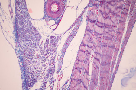
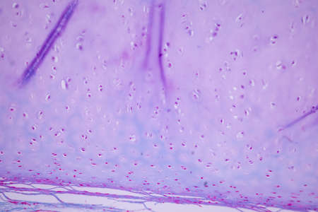
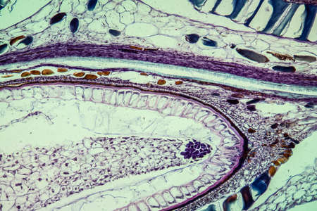
Cross section of human muscle under microscope view for education in laboratory.
Коллекция по умолчанию
Коллекция по умолчанию
Создать новую
abstraction background. red explosion circle on black background
Коллекция по умолчанию
Коллекция по умолчанию
Создать новую
Tissue of Small intestine (Duodenum) and Vermiform appendix Human under the microscope in Lab.
Коллекция по умолчанию
Коллекция по умолчанию
Создать новую
Microscopic view of tissue with purple and pink staining, showing cellular structures and nuclei
Коллекция по умолчанию
Коллекция по умолчанию
Создать новую
Microscopic view of tissue section showing cellular structures and layers, stained for examination
Коллекция по умолчанию
Коллекция по умолчанию
Создать новую
Tongue Tissue with taste buds across 100x
Коллекция по умолчанию
Коллекция по умолчанию
Создать новую
Pathology and Histology Tissue of Mammals under microscope.
Коллекция по умолчанию
Коллекция по умолчанию
Создать новую
Showing Light micrograph of the Adrenal gland and Urinary bladder human under the microscope for education in the laboratory.
Коллекция по умолчанию
Коллекция по умолчанию
Создать новую
Breast cancer, light micrograph, photo under microscope
Коллекция по умолчанию
Коллекция по умолчанию
Создать новую
Condyloma acuminatum, also known as genital warts. Light micrograph, photo under microscope
Коллекция по умолчанию
Коллекция по умолчанию
Создать новую
Ovarian mucinous cystadenoma, a benign tumor of ovary, light micrograph, photo under microscope
Коллекция по умолчанию
Коллекция по умолчанию
Создать новую
Characteristics of Lichen, hyphae and Symbiotic algae under the microscope for education.
Коллекция по умолчанию
Коллекция по умолчанию
Создать новую
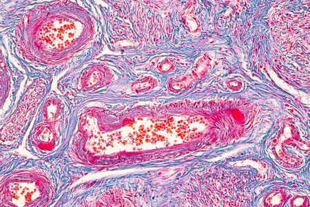
Connective tissue located at human outer ear, light micrograph. Hematoxylin and eosin stain
Коллекция по умолчанию
Коллекция по умолчанию
Создать новую
Characteristics of fungi living in wood as a group, are polyphyletic under the microscope for education.
Коллекция по умолчанию
Коллекция по умолчанию
Создать новую
Cross-section through the lichen symbiote body 100x
Коллекция по умолчанию
Коллекция по умолчанию
Создать новую

Showing Light micrograph of the Trachea, Thymus, Parathyroid gland and Tonsil human under the microscope for education in the laboratory.
Коллекция по умолчанию
Коллекция по умолчанию
Создать новую
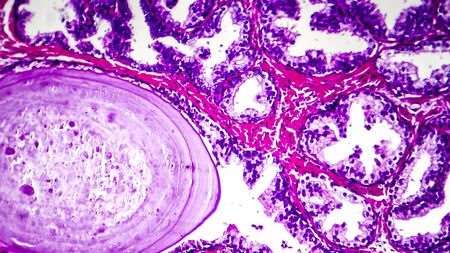
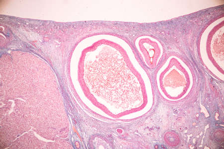
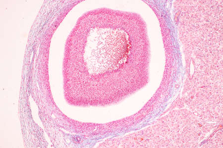
Benign prostatic hyperplasia. Micrograph shows dilated glands, papillary projections inside the lumen of the glands, cystic dilatation with accumulation of secretory material. Photo under microscope
Коллекция по умолчанию
Коллекция по умолчанию
Создать новую
Histopathology of human under microscope view for education in laboratory.
Коллекция по умолчанию
Коллекция по умолчанию
Создать новую
Microscopic Anatomy of Lycoperdon Bovista Puffball Gleys and Spore Structure
Коллекция по умолчанию
Коллекция по умолчанию
Создать новую
Leech cross section showing internal anatomical structures stained
Коллекция по умолчанию
Коллекция по умолчанию
Создать новую
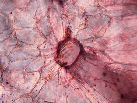
Microscopic view of stained carrot root cells. Cross section. Optical compound microscope. Brightfield. Objective 40x.
Коллекция по умолчанию
Коллекция по умолчанию
Создать новую

Education anatomy and Histological sample of Human under the microscope.
Коллекция по умолчанию
Коллекция по умолчанию
Создать новую
Characteristics of Lichen, hyphae and Symbiotic algae under the microscope for education.
Коллекция по умолчанию
Коллекция по умолчанию
Создать новую

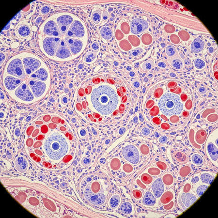
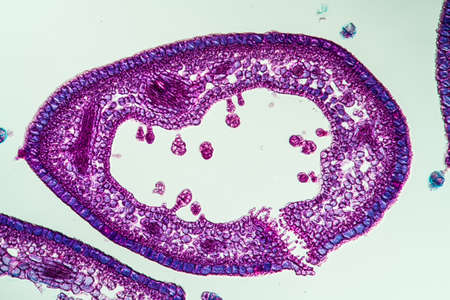
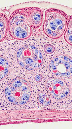
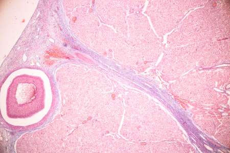
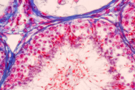
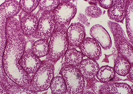
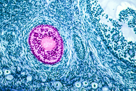
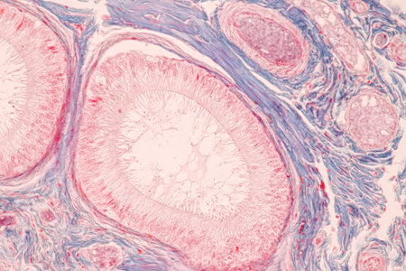
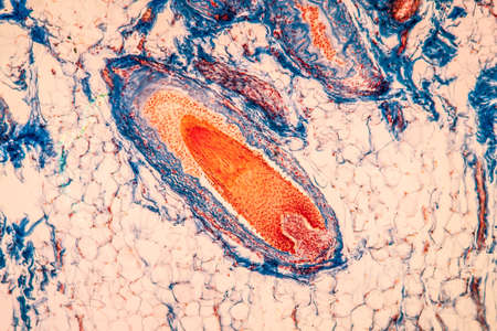
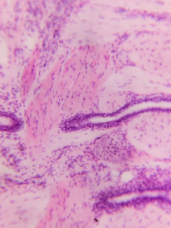
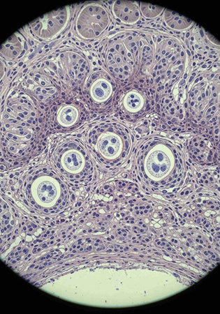
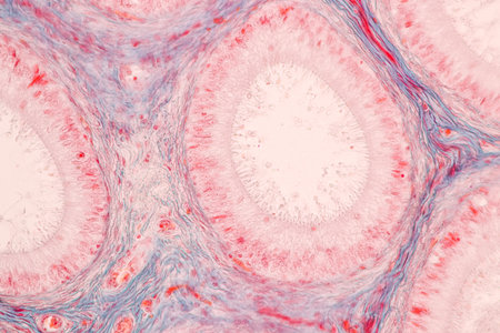
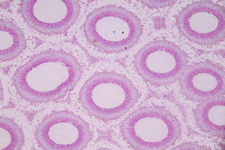
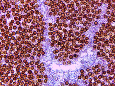
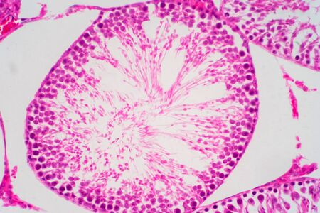
Tubules of sperm in epididymis
Коллекция по умолчанию
Коллекция по умолчанию
Создать новую
Characteristics of Lichen, hyphae and Symbiotic algae under the microscope for education.
Коллекция по умолчанию
Коллекция по умолчанию
Создать новую
Anatomy and Histological Ovary, Testis and Sperm human cells under microscope.
Коллекция по умолчанию
Коллекция по умолчанию
Создать новую
Anatomy and Histological Ovary, Testis and Sperm human cells under microscope.
Коллекция по умолчанию
Коллекция по умолчанию
Создать новую
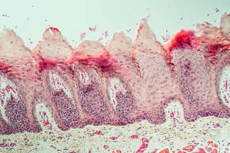
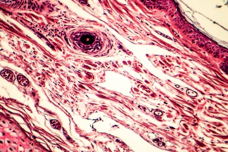
Cross section human skin head under microscope view for education histology. Histological for human physiology.
Коллекция по умолчанию
Коллекция по умолчанию
Создать новую
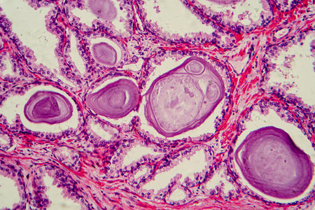
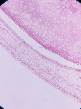
Elastic cartilage of human outer ear, light micrograph
Коллекция по умолчанию
Коллекция по умолчанию
Создать новую

Ovarian cancer, light micrograph, photo under microscope. Photograph shows a fragment of a cancerous tumor in the female ovary. Selective focus
Коллекция по умолчанию
Коллекция по умолчанию
Создать новую
A longitudinal section of human spinal ganglion cells under the microscope.
Коллекция по умолчанию
Коллекция по умолчанию
Создать новую

Cross-section through the lichen symbiote body 100x
Коллекция по умолчанию
Коллекция по умолчанию
Создать новую

Human endometrium. Proliferative phase. Several tubular endometrial glands are cross sectioned showing their simple columnar epithelium. Among them, the uterine stroma shows many cells and blood vessels.
Коллекция по умолчанию
Коллекция по умолчанию
Создать новую
Human pancreas under microscope view. Histological sample for human biology.
Коллекция по умолчанию
Коллекция по умолчанию
Создать новую
Structure of human cell under microscope view. 3D illustration.
Коллекция по умолчанию
Коллекция по умолчанию
Создать новую
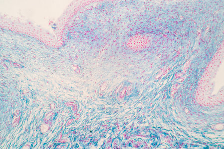
Human skin with a scabies infection, Sarcoptes scabiei, under the microscope and with coloration.
Коллекция по умолчанию
Коллекция по умолчанию
Создать новую
Pancreas cancer cell under microscope view for medical education.
Коллекция по умолчанию
Коллекция по умолчанию
Создать новую

micrograph plant tissue, stem of pumpkin
Коллекция по умолчанию
Коллекция по умолчанию
Создать новую
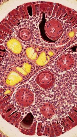
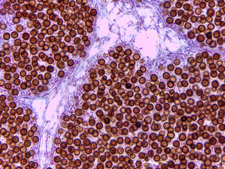
Human sperm in the testis morphology under microscope. Micrograph showing spermatozoon for pathology education.
Коллекция по умолчанию
Коллекция по умолчанию
Создать новую
Pathology and Histology Tissue of Mammals under microscope.
Коллекция по умолчанию
Коллекция по умолчанию
Создать новую

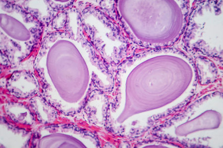
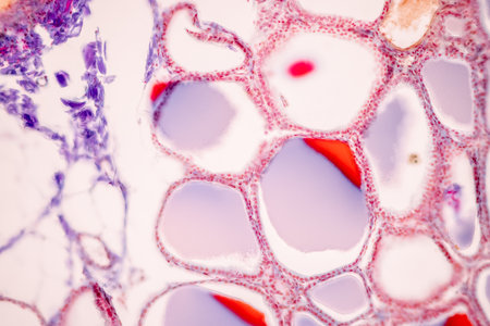
Endemic goiter, light micrograph, abnormal enlargement of the thyroid gland due to dietary iodine deficiency. Photomicrograph shows follicles of varying size, abundant colloid, lymphocytic infiltrate
Коллекция по умолчанию
Коллекция по умолчанию
Создать новую
Abstract background of acrylic. Colourful rpaint dissolve
Коллекция по умолчанию
Коллекция по умолчанию
Создать новую
Legion-Media
Создайте свои проекты на основе качественных стоковых фотографий и видео.


Copyright © Legion-Media.